Category:Lungs
English: Lungs
Esperanto: Pulmoj
Polski: Płuca
Slovenščina: Pljuča
Suomi: Keuhkot
Walon : Peumons
essential respiration organ in many air-breathing animals  Pulmons humans (German) | |||||
| Upload media | |||||
| Pronunciation audio | ⓘ | ||||
|---|---|---|---|---|---|
| Instance of |
| ||||
| Subclass of |
| ||||
| Part of | |||||
| Different from | |||||
| |||||
Subcategories
This category has the following 25 subcategories, out of 25 total.
- Videos of lungs (10 F)
A
- Airway basal cells (21 F)
B
- Bronchiolar exocrine cells (11 F)
- Brush cells (8 F)
C
- Club cells (10 F)
D
- Deuterosomal cells (5 F)
- Deuterosomal cels (empty)
H
I
- Ionocytes (6 F)
L
- Lung stem cells (4 F)
P
- Pneumocytes (16 F)
- Pulmonary imaging (7 F)
R
- Respiratory failure (9 F)
Media in category "Lungs"
The following 74 files are in this category, out of 74 total.
-
1 IPF Lung Sound.wav 31 s; 5.68 MB
-
 1D53EE7 image.jpg 5,712 × 4,284; 2.62 MB
-
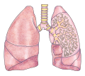
 202008 lung detailed.svg 512 × 224; 2.85 MB
-
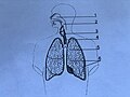
 202008 lung.svg 512 × 453; 1.13 MB
-
 6 Laktanya Street, 2020 Óbuda.jpg 2,272 × 1,704; 1,000 KB
-
 A Three Pig Lungs.jpg 3,888 × 5,184; 8.68 MB
-
 Air curtain sign of a normal lung as shown on ultrasound in sagittal view.png 1,024 × 698; 188 KB
-
 Airway epithelial differentiation pathways.png 3,233 × 3,491; 2.12 MB
-
 Alpha enolase graph.png 425 × 339; 60 KB
-
 Alveolar duct2.JPG 2,816 × 2,112; 2.08 MB
-
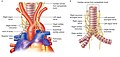 Anatomytool Dually innervated heart and lungs English.jpg 810 × 388; 51 KB
-
 Anatomytool Lungs and chest wall English.jpg 770 × 630; 122 KB
-
 Annie Cattrell – Capacity.jpg 1,200 × 900; 468 KB
-
 Birikak.png 579 × 612; 173 KB
-
 Birikaren zokoguneak 1.png 673 × 638; 155 KB
-
 Buffalo medical journal (1913) (14764087915).jpg 1,411 × 1,866; 745 KB
-
 BỆNH VIỆN THẬN HÀ NỘI.jpg 142 × 142; 15 KB
-
 Casts of lungs, Marco resin, 1951 (24226157742).jpg 800 × 1,237; 478 KB
-
 COR-2-STND-CHEST-LUNGS.jpg 1,173 × 895; 257 KB
-
 CT-Low-Dose-1.25-Lung-Calcified-Nodule.jpg 1,920 × 1,080; 619 KB
-
CT-Low-Dose-2.5-LUNG.ogg 36 s, 1,490 × 896; 10.87 MB
-
 CT-Standard-Dose-2.50-Lung-Calcified-Nodule.jpg 1,920 × 1,080; 579 KB
-
 CT-Thorax-5.0-B70f-Lungs.jpg 1,024 × 1,024; 531 KB
-
De-Lunge.ogg 1.5 s; 15 KB
-

-

-

-
En-us-lungs.ogg 1.2 s; 14 KB
-

-

-
-
 FACT- Vaccines against pneumonia DO NOT protect against the COVID-19 virus.png 1,024 × 512; 144 KB
-
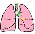 Ghonfoküsü.jpg 290 × 285; 20 KB
-
 Guts - Flickr - striatic.jpg 987 × 1,316; 952 KB
-

-

-

-
 Image of lungs and liver.jpg 4,000 × 1,800; 1.91 MB
-
 Image saturation due to overexposure on chest X-ray.jpg 957 × 968; 166 KB
-
 Journal of morphology (1893) (14596056270).jpg 3,262 × 2,376; 649 KB
-
 Key ILC2 interactions in lung development, inflammation, and fibrosis.png 3,147 × 2,841; 2.6 MB
-

-
Low dose high resolution chest CT (HRCT).ogg 12 s, 1,920 × 1,080; 15.59 MB
-
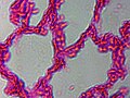 Lung cells.jpg 640 × 480; 74 KB
-
 Lungs in museum.jpg 588 × 861; 190 KB
-
MP1 Pulmonology.webm 5 min 1 s, 1,280 × 720; 217.94 MB
-
 Obstrukce vs restrikce.jpg 800 × 1,131; 57 KB
-

-
 Overview of the nasal epithelium, showing various cell types (black arrow).png 4,042 × 2,090; 2.15 MB
-
 Peripheral localization and distinct morphology of Gr1+ cells present in necrotic lungs.png 2,049 × 1,542; 2.95 MB
-
 PIV setup.png 593 × 591; 189 KB
-
 Pneumonia forming around bronchioles.png 350 × 310; 259 KB
-
 Potential role of PNECs in brain diseases.jpg 4,212 × 2,343; 481 KB
-

-
 Pulmão e coraçã.jpg 683 × 754; 140 KB
-
 Role of PNECs in physiological and pathological conditions in lung.jpg 4,095 × 2,946; 523 KB
-

-

-
 Schematic representation of epithelial cells lining the airways.png 3,741 × 1,688; 1.14 MB
-
 Schematic representation of secretory cell (SC) differentiation programs.png 3,119 × 2,090; 373 KB
-
 Smoker's lungs.jpg 558 × 802; 195 KB
-
 Sopa de letras pulmones.png 487 × 583; 63 KB
-
Standard dose high resolution chest CT (HRCT).ogg 27 s, 1,920 × 1,080; 33.01 MB
-
 Structure of the airway epithelium.png 850 × 402; 293 KB
-

-

-
 Three anatomical figures from Tibet Wellcome V0036134.jpg 2,691 × 3,215; 3.36 MB
-
 Timeline of PNECs research.jpg 4,327 × 3,495; 496 KB
-

-
 TRM niches in the in the lung.jpg 758 × 703; 459 KB
-
 UOEOxygen.png 256 × 256; 9 KB
-
 Wakan Sansai Zue - Hai.jpg 300 × 358; 43 KB
-
 Эффективный циркуляционный объем31.jpg 1,268 × 1,938; 492 KB
-
 ఊపిరితిత్తులు, శ్వాసనాళ శాఖలు (Lungs and Bronchial tree).png 2,342 × 2,448; 2.76 MB


_(14764087915).jpg)

.jpg)


_cells_on_decellularized_lung_scaffolds_resembles_in_situ_epithelial_differentiation_of_native_mouse_airways.png)


.png)


_cells.webp)


_(14596056270).jpg)

.jpg)


.png)


_differentiation_programs.png)


_cells_seeded_on_decellularized_lung_scaffolds_do_not_differentiate_into_proximal_airway_cells.webp)


.png)
_cells._Cropped.jpg)
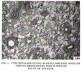
_centrosome_highlighting_the_9_%2B_0_arrangement_of_triplet_microtubules_(box)_and_(b)_a_cilium_showing_9_%2B_2_arrangement_(motile_cilium)_of_doublet_microtubules_(box.png)