Category:Coelom
main body cavity in most animals  | |||||
| Upload media | |||||
| Subclass of |
| ||||
|---|---|---|---|---|---|
| Different from | |||||
| |||||
Media in category "Coelom"
The following 57 files are in this category, out of 57 total.
-
 Abatus cordatus Long-spined juvenile (J2) (01).jpg 1,032 × 1,402; 1.43 MB
-
 Abatus cordatus Long-spined juvenile (J2).jpg 879 × 1,080; 781 KB
-
 Abatus cordatus Post-gastrular stage 100 days after fertilization.jpg 1,025 × 1,566; 1.41 MB
-
 Abatus cordatus Post-gastrular stage.jpg 930 × 1,284; 1.04 MB
-
 Abatus cordatus Short-spined juvenile (J1).jpg 1,711 × 739; 1.05 MB
-

-
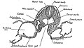
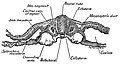
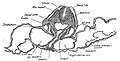
 Amphioxus Origin of the mesoderm.jpg 1,802 × 756; 793 KB
-
 Annelid redone w white background.svg 3,732 × 2,760; 7.65 MB
-
-

-
-
-
-
-

-

-
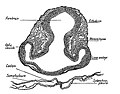
 Aves Transverse sections through embryo fifth day after incubation.jpg 688 × 755; 357 KB
-

-
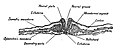
 Branchiostoma formation of the neural tube, notochord, mesoderm, and coelom.jpg 1,172 × 857; 562 KB
-
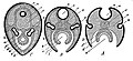
 Coelomate 01-2.png 250 × 250; 20 KB
-
 Coelomate 01.png 250 × 250; 12 KB
-
 Development of the amnion and allantois.jpg 676 × 902; 1.05 MB
-

-

-
 Early human embryo (01).jpg 675 × 563; 209 KB
-
 Early human embryo (02).jpg 915 × 432; 257 KB
-
 Early human embryo.jpg 761 × 738; 272 KB
-
 EB1911 Peripatus - Series of Embryos.jpg 1,011 × 723; 218 KB
-
 Ectopleura crocea. blastula (left) morula (right).jpg 1,074 × 825; 557 KB
-
 Figure 27 02 05.jpg 1,117 × 738; 593 KB
-
 Formation of the Umbilical Region. human embryo, 1.7 mm. long.jpg 1,065 × 857; 974 KB
-
 Formation of the Umbilicus and Allantois. human embryo, 0.7 mm. long..jpg 1,152 × 803; 970 KB
-
 Formation- of the Umbilicus in an Embryo 2.5 mm.jpg 788 × 837; 598 KB
-
 Human embryo Section of embryonic rudiment in Peters' ovum (first week).jpg 1,141 × 857; 540 KB
-

-
 Human embryo Transverse Section.jpg 932 × 792; 477 KB
-

-
 Human- Embryo, about 5 mm. long.jpg 825 × 846; 809 KB
-
 Kirkes' handbook of physiology (1907) (14769687492).jpg 1,091 × 918; 435 KB
-
 Location of the intermediate mesoderm nephrogenic cord..jpg 600 × 227; 58 KB
-
-
 Origin of Vertebrates Fig 157.png 1,282 × 552; 30 KB
-
 Sagittal Section of Human Zygote.jpg 817 × 684; 653 KB
-

-
 Stages of mammals embryos.jpg 976 × 1,102; 849 KB
-

-

-
 Umbilical Abnormal Structures (02).jpg 778 × 562; 393 KB
-
 Umbilical Cord of a Human Embryo 12.5 mm. in length.jpg 1,001 × 1,445; 1.15 MB
-
 Umbilical Cord of a Human Embryo 18 mm. in length.jpg 601 × 779; 537 KB
-
 Umbilical Region in a Human Embryo 12 cm. in Length.jpg 707 × 707; 585 KB
-
 Umbilical Region in an Embryo 7 mm. in length.jpg 882 × 713; 492 KB
-
 Umbilical Region of a Fetus at Term. Abnormal.jpg 613 × 1,116; 498 KB
-

-
 Umbilical Region of a Human Embryo 5.2 cm. in Length.jpg 864 × 945; 763 KB
-
-
 Vetebrateembryo.svg 716 × 604; 197 KB
_(01).jpg)
.jpg)


.jpg)


,_myotome_(muscle-producer),_and_sclerotome_(skeleton-producer).jpg)
.jpg)
.jpg)


_morula_(right).jpg)


.jpg)

_(14769687492).jpg)


.jpg)


