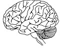
Category:Brain
organ that serves as the center of the nervous system in all vertebrate and most invertebrate animals  | |||||
| Upload media | |||||
| Spoken text audio | ⓘ | ||||
|---|---|---|---|---|---|
| Pronunciation audio | ⓘ | ||||
| Instance of |
| ||||
| Subclass of |
| ||||
| Part of |
| ||||
| Connects with |
| ||||
| Has part(s) | |||||
| Different from | |||||
| |||||
English: NOTE: Anatomy files should be categorized by species!.
Français : Cerveau
Türkçe: Beyin
Subcategories
This category has the following 31 subcategories, out of 31 total.
*
- Audio files of brain (41 F)
5
- 52 brain facts (18 F)
A
B
C
D
G
- Gut–brain axis (15 F)
H
L
M
- Emeran Mayer (2 F)
N
O
R
S
T
V
- Brain vital signs (1 F)
Media in category "Brain"
The following 200 files are in this category, out of 234 total.
(previous page) (next page)-
 1 - copia.png 600 × 600; 416 KB
-
 1907 image of a brain (Labour and Childhood).png 1,233 × 958; 1 MB
-
 2009 cerveau corail 2D.JPG 1,018 × 881; 65 KB
-
 2010-3-15 rGFAP 1-4000 1-200 Hip 20x(4).tif 2,040 × 1,536; 8.99 MB
-
 2017 First Amparo Workshop 019.jpg 4,437 × 3,327; 2.04 MB
-
 21318-0550x0475 (2).jpg 421 × 364; 75 KB
-
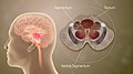 3D Medical Animation Mid-Brain Different Parts.jpg 1,920 × 1,080; 1.27 MB
-
 Teste testando.jpg 527 × 550; 29 KB
-
 ACC in rat brain.png 443 × 312; 92 KB
-
 ADHD.jpg 1,920 × 1,080; 1.39 MB
-
 African brain.png 503 × 264; 98 KB
-
 Aires cérébrales chargées de l'équilibre.png 1,002 × 499; 27 KB
-
 Aires cérébrales de la motricité volontaire.jpg 456 × 428; 43 KB
-
 Allo-endo.tif 1,710 × 791; 3.89 MB
-
 Ancient Meitei character "KOK" (ꯀ) represents as well as means "brain" or "head".jpg 5,044 × 2,158; 696 KB
-

-
 Association Cortices.png 711 × 400; 29 KB
-
 Astrocyte et neurone.png 850 × 1,057; 260 KB
-

-
 Aupegi nerbioa 1.png 1,200 × 1,429; 706 KB
-
 Bbb cells.png 1,375 × 675; 348 KB
-
 Blood brain barriere cells.png 828 × 639; 295 KB
-
 Blood vessels brain english.jpg 868 × 537; 126 KB
-
 Blood–Brain Barrier Breakdown in Alzheimer’s Disease.png 1,375 × 768; 464 KB
-
 Blue Brain 3 (Small).JPG 1,400 × 285; 56 KB
-
 Blue Brain.jpg 640 × 298; 32 KB
-
 BMR TEE age % Brain energy.png 3,208 × 1,949; 34 KB
-
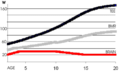 BMR TEE brain energy change with age..png 2,964 × 1,808; 29 KB
-
 Brain cell(s) (954701212).jpg 3,888 × 2,592; 2.46 MB
-
 Brain Cells (40522751690).png 556 × 615; 653 KB
-
 Brain Chip (40529717123).jpg 591 × 592; 48 KB
-
 Brain Chip (40529718453).jpg 960 × 882; 265 KB
-
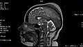
 Brain CT scan.jpg 900 × 1,122; 377 KB
-
 Brain infograph.png 4,500 × 3,000; 14.93 MB
-
 Brain Maps.JPG 1,915 × 1,033; 340 KB
-
 Brain regions in memory formation updated.jpg 1,284 × 889; 122 KB
-
 Brain Sensor.jpg 3,534 × 2,832; 3.02 MB
-

-
 Brain Study of Frontal Lobe.png 1,080 × 1,080; 741 KB
-
 Brain Tissue Cells (33272648413).png 556 × 310; 326 KB
-
 Brain Using Energy.jpg 2,624 × 1,348; 1,018 KB
-
 Brain ventricular system.stl 5,120 × 2,880; 1.81 MB
-
 Brainiac.JPG 3,168 × 4,752; 3.97 MB
-
 Brainpicture.png 1,200 × 1,200; 403 KB
-
 Brains Season 2 Poster.jpg 2,700 × 3,600; 6.91 MB
-
 Brainstem divisions.png 623 × 472; 36 KB
-
 BrainSTree.pdf 1,500 × 1,125; 794 KB
-
 BrainWaves Brain Waves.png 700 × 450; 102 KB
-
 Brainwaves Representation.png 700 × 450; 179 KB
-
 Brand in Brain.png 1,168 × 480; 537 KB
-
 Breakdown in Blood–Brain Barrier in Alzheimer’s Disease.png 649 × 515; 240 KB
-
 Brisa Alfaro, 32 year old Pons Stroke and Locked In Syndrome Survivor.jpg 2,448 × 3,264; 1.98 MB
-
 BROCA'S.jpg 1,728 × 2,304; 483 KB
-
 Brodmann's areas of human brain.jpg 1,913 × 1,080; 407 KB
-
 C2orf72 Orthologs List.png 807 × 616; 305 KB
-
 Captura de pantalla 2018-05-10 a las 11.52.58.png 275 × 297; 92 KB
-
 Cells-09-00439-g001.jpg 800 × 395; 49 KB
-
 Cells-09-00439-g003.jpg 702 × 550; 69 KB
-
 Cells-09-00439-g004.jpg 789 × 593; 75 KB
-
 Cerebral Folate Deficiency - Cerebral CT-scan at 4 years old.png 2,986 × 1,556; 644 KB
-
 Cerebro humano.jpg 4,000 × 3,000; 3.64 MB
-
 Cervello.png 200 × 160; 45 KB
-
 Charles Bell Anatomy of the Brain, c. 1802 (3138247450).jpg 551 × 495; 92 KB
-
 Comparative evolution of the striatum and pallium in vertebrates.png 2,902 × 1,370; 2.45 MB
-
 Connectivity matrix.jpg 852 × 807; 96 KB
-
 Connectomics.png 1,103 × 976; 1.37 MB
-
 Constuddivis.png 431 × 472; 6 KB
-
 Constudoverbrain3.svg 1,063 × 709; 24 KB
-
 Coronal section of non-functioning pituitary adenoma.jpg 3,456 × 4,608; 6.36 MB
-
 Coronal view of the MRI brain with double inversion sequence.png 3,456 × 4,608; 25.65 MB
-
 CoronalBrain.jpg 1,280 × 1,920; 871 KB
-
 Coup injury.jpg 1,920 × 1,080; 676 KB
-
-
 CT brain of posterior interhemispheric bleed.jpg 512 × 512; 126 KB
-
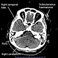
 CT brain showing haematoma on skull vertex.jpg 3,456 × 4,608; 7.77 MB
-

-
 CT scan of left small parietal bleed of brain.jpg 512 × 512; 131 KB
-
 CT scan of the brain showing recent right basal ganglia bleed.jpg 512 × 512; 139 KB
-
-
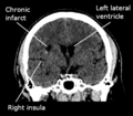
 CT scan showing chronic infarct at the left high parietal region.jpg 3,456 × 4,608; 11.18 MB
-
 CT scan showing chronic Infarct at the right corona radiata.png 413 × 361; 111 KB
-
 CT scan showing Pacchionian granulation at left sigmoid sinus.jpg 512 × 512; 128 KB
-
 Curva PIC.jpg 467 × 425; 46 KB
-
 Default Mode Network Connectivity.png 3,491 × 7,927; 32.23 MB
-
 Development Sequences of the Human Mind.jpg 2,007 × 1,226; 369 KB
-
 DGC dispersion.jpg 1,000 × 413; 128 KB
-
 Dissociative identity disorder neuroscience brain imaging (no description).png 1,391 × 631; 342 KB
-
 Dr. Edith Klemperer Patent for Luminous Brain Model, 1934 Cutout.png 1,212 × 1,272; 99 KB
-
 Dr. Edith Klemperer Patent for Luminous Brain Model, 1934.png 407 × 615; 116 KB
-
 Dual Process Brain.png 1,040 × 972; 323 KB
-
 Dyslexia.jpg 1,920 × 1,080; 812 KB
-
 Editorial on gravity in the brain by J. Howard Jaster, MD—final gallery proof, in June 2021.pdf 1,275 × 1,650, 4 pages; 439 KB
-

-

-
 Egad isoforms rat brain diff ages.png 1,993 × 3,932; 855 KB
-
 Electron micrograph of a Fractone.jpg 3,662 × 3,472; 2.37 MB
-
 En hjärnas födelse och död.jpg 502 × 720; 122 KB
-
 Esquema accion hormonal.jpg 795 × 517; 71 KB
-
 Example of a Nissl stain of an avian brain.tif 963 × 964; 3.73 MB
-
 Faces file upload of Corey’s artwork 03.jpg 890 × 716; 284 KB
-
 Facts abpout the Amygdala!.jpg 1,093 × 2,362; 307 KB
-
 Fantoom.PNG 412 × 273; 7 KB
-
 Fast-Biopsychology.pdf 2,000 × 1,125, 2 pages; 256 KB
-

-
-
 Figure charge 238 273 mod.png 1,920 × 1,056; 2.14 MB
-
 Fneur-10-00574-g001.jpg 765 × 543; 277 KB
-
 Fnins-14-00754-g002.jpg 2,250 × 1,807; 401 KB
-
 Folic acid metabolism and 5-MTHF transport RUS.png 3,721 × 1,266; 3.43 MB
-
 Freqenztypen.png 867 × 233; 33 KB
-
 Frontallobes.png 339 × 226; 30 KB
-
 Gad65 and gad67 in rat brain diff ages.png 2,807 × 1,821; 925 KB
-
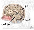
 Gehirn beschriftet.svg 512 × 351; 450 KB
-
 GH controle.png 1,218 × 981; 374 KB
-
 Gut-brain-skin axis.jpg 550 × 358; 31 KB
-
 Hall of Human Life Museum Science Boston.jpg 3,240 × 4,320; 2.24 MB
-
 HD@DH.nrw differenziertes Lernen.svg 512 × 420; 13 KB
-
 HD@DH.nrw Gehirn mit verschieden hervorgehobenen Bereichen.svg 512 × 290; 61 KB
-
 HD@DH.nrw Gehirn.svg 512 × 451; 6 KB
-
 HD@DH.nrw Lerndefizite.svg 512 × 503; 12 KB
-
 HD@DH.nrw Lerntempo.svg 512 × 468; 10 KB
-
 HD@DH.nrw medial vielfältiges Lernen.svg 512 × 464; 17 KB
-
 HD@DH.nrw Robotergehirn.svg 512 × 429; 4 KB
-
 Head involution. Aphidoletes aphidimyza.png 850 × 584; 597 KB
-
 HFO wiki.jpg 1,534 × 684; 79 KB
-
 Hippocampus scheme.jpg 1,128 × 658; 161 KB
-
 Homologous Regions between Bird and Human Brains.png 800 × 450; 277 KB
-
 HPA RNA-Seq normal tissues C2Orf72 Expression Profile July 16 2022.png 2,241 × 840; 60 KB
-
 Hypothalamus (1).pdf 3,000 × 1,687, 6 pages; 525 KB
-
 Hypothalamus Canva PSY 1000.png 800 × 2,000; 361 KB
-
 Hypothalamus Infographic.png 1,728 × 2,304; 1.23 MB
-
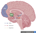
 HYPOTHALAMUS.png 800 × 600; 324 KB
-
 I-TASSER C2Orf72 Summer 2021 structure prediction.png 977 × 1,028; 473 KB
-
 Inferior Temporal Lobe Cortex.pdf 1,250 × 3,125; 1.19 MB
-
 Isihlahla samaganandoda 'Pear tree'.jpg 1,536 × 2,048; 1.12 MB
-
 Isshūkan Friends. logo.png 619 × 129; 51 KB
-
 Jaime Lara 2023 PMID 36409650 Figure 8.jpg 750 × 584; 135 KB
-
 Jill Bolte Taylor - observing a stroke from within.jpg 1,987 × 2,271; 1.15 MB
-
 Journal.pone.0039279.g002.png 4,662 × 1,465; 119 KB
-
 Journal.pone.0039279.g003.png 4,654 × 1,486; 112 KB
-
 Journal.pone.0039279.g004.png 5,236 × 2,217; 266 KB
-
 Journal.pone.0039279.g006.png 5,234 × 2,217; 267 KB
-
 Journal.pone.0052528.g003.png 1,910 × 1,381; 359 KB
-
 Journal.pone.0057573.g005.png 909 × 588; 550 KB
-
 L'Abbé Desmonceaux. Line engraving by B. A. Nicollet after L Wellcome V0001565.jpg 2,224 × 3,276; 3.86 MB
-
 Labeled cichlid forebrain diagram with shh expression.jpg 702 × 580; 111 KB
-
 Laboratory of neural engineering (2692819292).jpg 2,048 × 1,536; 604 KB
-
 Lateral View of Human Brain (Black and White).jpg 1,542 × 1,252; 507 KB
-
 Left frontal extra-axial lesion causing vasogenic odema and midline shift to the right.jpg 3,456 × 4,608; 7.82 MB
-
 Lobulo linbikoa.jpg 3,769 × 2,126; 1.12 MB
-
 Lobulo linbikoaren atalak (erdialdeko ikuspegia).jpg 3,891 × 2,059; 1.21 MB
-
 Maquina 23.jpg 750 × 562; 72 KB
-
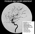 MCA angio lateral-hu.jpg 1,156 × 1,128; 544 KB
-
 Meningioma of the sagittal sinus isolated.jpg 2,000 × 1,146; 396 KB
-
 Mesocorticolimbic Circuit.png 683 × 622; 204 KB
-
 Meyeline.png 1,309 × 431; 532 KB
-
 Microzone.svg 648 × 451; 23 KB
-
 Mindfulness brain.jpg 1,920 × 1,239; 1,013 KB
-
 MLIS STRING 2.png 2,247 × 1,451; 1.25 MB
-
 MLIS-STRING10.png 2,337 × 1,716; 1.52 MB
-

-

-
 Motor loop.png 1,024 × 768; 689 KB
-
 Motor Nerve Pathways Descending.png 720 × 1,200; 632 KB
-
 MRI brain of extra-axial lesion at right frontal lobe with perilesional oedema.png 6,912 × 4,608; 38.58 MB
-
 MRI Brain Septum Deviation.png 766 × 947; 509 KB
-
 MRI Capas.png 601 × 367; 235 KB
-
 Myelin Sheath.jpg 800 × 2,000; 233 KB
-
 Máquina para la Tomografía por Emisión de Positrones.jpg 176 × 128; 4 KB
-
 Neocortex séma.jpg 673 × 330; 70 KB
-
 Neural pathway diagram ge.svg 512 × 425; 48 KB
-
 OpenBrainGraph-withLegend.png 1,126 × 625; 100 KB
-
 Origins of Motivation in Primates.jpg 1,965 × 1,239; 451 KB
-
 Parallel-fibers.png 389 × 465; 64 KB
-
 Particulates exposure and increased risk of neurodegeneration.jpg 800 × 330; 75 KB
-
 Parvo magno.PNG 749 × 696; 33 KB
-
 Perfiles de tipos de células.png 2,100 × 1,915; 2.26 MB
-
 Plasticity Specific Pathways it.png 371 × 555; 70 KB
-
-
-
Prediction-of-Biological-Motion-Perception-Performance-from-Intrinsic-Brain-Network-Regional-Video3.ogv 5.0 s, 1,280 × 720; 107 KB
-
Prediction-of-Biological-Motion-Perception-Performance-from-Intrinsic-Brain-Network-Regional-Video4.ogv 5.0 s, 1,280 × 720; 106 KB
-
 Prefrontal Cortex part of the brain function.png 1,728 × 2,304; 1.34 MB
-
 Psych cerebellum poster.jpg 3,344 × 2,508; 2.27 MB
-
 Psych204b DF Fig1.png 1,620 × 928; 914 KB
-
 PVH neurons of Long-Evans rat marked with retrograde tracer floro-gold.tif 1,280 × 1,024; 7.54 MB
-
 Retardo Interauricular.png 2,106 × 4,221; 816 KB
-
 Régulation cardiovasculaire centrale.png 1,262 × 575; 35 KB
-

-
 Series 001 (3-PL LOC).png 256 × 256; 65 KB
-
 Sopa de letras encefalo.png 448 × 547; 34 KB
-
 Specifieke kerngroepen in thalamus en hun functie.png 533 × 440; 16 KB
-
 STRING MLIS.jpg 1,342 × 938; 287 KB
-
 STRING MLIS.png 3,078 × 2,086; 2.62 MB
-
 Structure of the brain.jpg 3,024 × 4,032; 2.9 MB
-
 Subcortical atlas regions.pdf 1,604 × 1,289; 4.61 MB
-
 Substantia Nigra Pars Reticula Deep Brain Stimulation.png 1,728 × 2,304; 3 MB
-
 Superior View of the Brain.jpg 521 × 457; 56 KB
-
 Synaptic glomerule from the red nucleus of the kat's brain.png 564 × 494; 380 KB
-


.png)


.jpg)


_represents_as_well_as_means_%22brain%22_or_%22head%22.jpg)
_-_meaning_as_well_as_representing_%22head%22_or_%22brain%22.jpg)


_(954701212).jpg)
.png)
.jpg)
.jpg)


.png)


.jpg)


.png)


_(21001294879).jpg)
_(21188129365).jpg)


.jpg)
.jpg)


.jpg)


.png)


.JPG)