
Sınaq göstərişi ölçüsü: 482 × 600 piksel. Digər ölçülər: 193 × 240 piksel | 386 × 480 piksel | 617 × 768 piksel | 823 × 1.024 piksel | 1.800 × 2.239 piksel.
Faylın orijinalı (1.800 × 2.239 piksel, fayl həcmi: 1,33 MB, MIME növü: image/jpeg)
Bu fayl Vikianbarda yerləşir. Açıqlama səhifəsindəki məlumatlar aşağıda göstərilib. Vikianbar azad lisenziyalı media anbarıdır. Siz də töhfə verə bilərsiniz. |
Xülasə
| İzah |
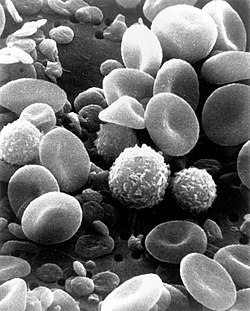
English: This is a scanning electron microscope image from normal circulating human blood. One can see red blood cells, several white blood cells including lymphocytes, a monocyte, a neutrophil, and many small disc-shaped platelets. Red cells are nonnucleated and contain hemoglobin, an important protein that contains iron and allows the cell to carry oxygen to other parts of the body. They also carry carbon dioxide away from peripheral tissue to the lungs where it can be exhaled. The infection-fighting white blood cells are classified in two main groups: granular and agranular. All blood cells are formed in the bone marrow. There are two types of agranulocytes: lymphocytes, which fight disease by producing antibodies and thus destroying foreign material, and monocytes. Platelets are tiny cells formed in bone marrow and are necessary for blood clotting. Type: Black & White Print Русский: Это изображение нормально циркулирующей крови человека получено с помощью сканирующего электронного микроскопа. Можно видеть красные кровяные тельца, несколько белых клеток крови (в их числе лимфоциты, моноциты, нейтрофилы) и множество мелких дискообразных пластинок. Красные кровяные тельца содержат гемоглобин — важный белок, который содержит железо и позволяет клетке переносить кислород к другим частям тела. Также они переносят углекислый газ от периферических тканей в лёгкие, где тот после газообмена может быть выдохнут. Лейкоциты борются с инфекциями и на две основные группы: гранулярные и агранулярные. Все клетки крови образуются в костном мозге. Есть два типа агранулоцитов: лимфоциты, которые борются с болезнью, производя антитела и тем самым разрушая чужеродный материал, и моноциты. Тромбоциты представляют собой крошечные клетки, образующиеся в костном мозге, и необходимы для свертывания крови. Тип фото: чёрно-белая печать. العربية : صورة بالمجهر الإلكتروني الماسح لدم الإنسان. يمكن للمرء أن يرى خلايا الدم الحمراء والعديد من خلايا الدم البيضاء بما في ذلك الخلايا الليمفاوية ووحيدات النوى والخلية المتعادلة والعديد من الصفائح الدموية الصغيرة ذات الشكل القرصي. |
||||||
| Tarix | Date Created: February 1982 | ||||||
| Mənbə | Image and description: National Cancer Institute | ||||||
| Müəllif | Bruce Wetzel (photographer). Harry Schaefer (photographer) | ||||||
| İcazə (Faylın təkrar istifadəsi) |
|
||||||
| Digər versiyalar |
Derivative works of this file: |
||||||
| Annotations | This image is annotated: View the annotations at Commons |
Faylın tarixçəsi
Faylın əvvəlki versiyasını görmək üçün gün/tarix bölməsindəki tarixlərə klikləyin.
| Tarix/Vaxt | Miniatür | Ölçülər | İstifadəçi | Şərh | |
|---|---|---|---|---|---|
| hal-hazırkı | 18:17, 3 fevral 2021 | | 1.800 × 2.239 (1,33 MB) | Tm | Reverted to version as of 20:27, 7 October 2006 (UTC) |
| 04:50, 10 noyabr 2020 |  | 1.800 × 2.239 (309 KB) | Ratmanz | Optimized. | |
| 20:27, 7 oktyabr 2006 |  | 1.800 × 2.239 (1,33 MB) | DO11.10 | ||
| 03:00, 4 oktyabr 2006 |  | 1.800 × 2.239 (989 KB) | DO11.10 | {{Information |Description=This is a scanning electron microscope image from normal circulating human blood. One can see red blood cells, several white blood cells including lymphocytes, a monocyte, a neutrophil, and many small disc-shaped platelets. Red | |
| 01:09, 4 oktyabr 2006 |  | 500 × 326 (36 KB) | DO11.10 | {{Information |Description= A three-dimensional ultrastructural image analysis of a T-lymphocyte (right), a platelet (center) and a red blood cell (left), using a Hitachi S-570 scanning electron microscope (SEM) equipped with a GW Backscatter Detector. |
Faylın istifadəsi
Aşağıdakı 2 səhifə bu faylı istifadə edir:
Faylın qlobal istifadəsi
Bu fayl aşağıdakı vikilərdə istifadə olunur:
- ar.wikipedia.org layihəsində istifadəsi
- ar.wikiversity.org layihəsində istifadəsi
- ast.wikipedia.org layihəsində istifadəsi
- as.wikipedia.org layihəsində istifadəsi
- ba.wikipedia.org layihəsində istifadəsi
- be-tarask.wikipedia.org layihəsində istifadəsi
- be.wikipedia.org layihəsində istifadəsi
- bg.wikipedia.org layihəsində istifadəsi
- bn.wikipedia.org layihəsində istifadəsi
- bn.wikibooks.org layihəsində istifadəsi
- bs.wikipedia.org layihəsində istifadəsi
- ca.wikipedia.org layihəsində istifadəsi
- ce.wikipedia.org layihəsində istifadəsi
- ckb.wikipedia.org layihəsində istifadəsi
- cs.wikipedia.org layihəsində istifadəsi
- cv.wikipedia.org layihəsində istifadəsi
- cy.wikipedia.org layihəsində istifadəsi
- de.wikipedia.org layihəsində istifadəsi
- de.wikibooks.org layihəsində istifadəsi
- dv.wikipedia.org layihəsində istifadəsi
- el.wikipedia.org layihəsində istifadəsi
- el.wiktionary.org layihəsində istifadəsi
- en.wikipedia.org layihəsində istifadəsi
Bu faylın qlobal istifadəsinə baxın.