Butun axtardiqlarinizi tapmaq ucun buraya: DAXIL OLUN
  Mp4 Mp3 Axtar Yukle
  Video Axtar Yukle
  Shekil Axtar Yukle
  Informasiya Melumat Axtar
  Hazir Inshalar Toplusu
  AZERI CHAT + Tanishliq
  1-11 Sinif Derslikler Yukle
  Saglamliq Tibbi Melumat
  Whatsapp Plus Yukle(Yeni)

  • Ana səhifə
  • Təsadüfi
  • Yaxınlıqdakılar
  • Daxil ol
  • Nizamlamalar
İndi ianə et Əgər Vikipediya sizin üçün faydalıdırsa, bu gün ianə edin.

Fayl:Apikal4D.gif

  • Fayl
  • Müzakirə
  • Vikianbar üzərindən baxış
  • Lokal izah əlavə et
  • Fayl
  • Faylın tarixçəsi
  • Faylın istifadəsi
  • Faylın qlobal istifadəsi
Fayl:Apikal4D.gif
Daha yüksək versiyası yoxdur.
Apikal4D.gif (636 × 432 piksel, fayl həcmi: 705 KB, MIME növü: image/gif, ilmələnib, 15 çərçivə, 0,6 s)
Bu fayl Vikianbarda yerləşir. Açıqlama səhifəsindəki məlumatlar aşağıda göstərilib.
Vikianbar azad lisenziyalı media anbarıdır. Siz də töhfə verə bilərsiniz.

Xülasə

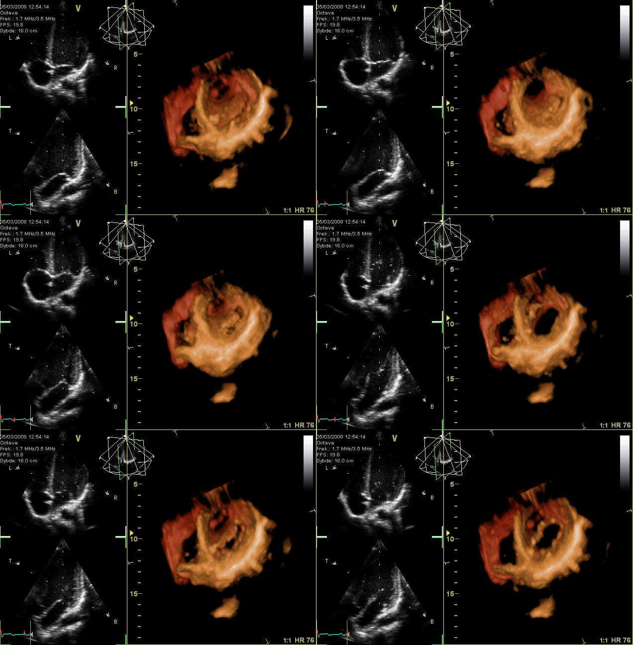
İzahApikal4D.gif
العربية: صُورةٌ مُتحرِّكة لِتخطيط صدى القلب؛ دائرةٌ كaهربائيَّة ثُلاثيَّة الأبعاد للقلب كما يبدو للناظر من الأعلى، حيثُ تمَّت إزالة الجُزء القمّي من البطينين بينما يظهر الصمَّام التاجي واضحًا للعيان. لا يظهر الصمَّامان الأبهر وثُلاثيّ الشرف بشكلٍ واضح نتيجة ضياع البيانات الخاصَّة بهذا التصوير، بينما تظهر الفتحات. يظهرُ على اليسار منظران ثُنائيَّا الأبعاد مأخوذان بناءً على بيانات الرسم ثُلاثي الأبعاد. يُفسِّرُ هذا الرسم الصُورة المُتحرِّكة.
English: GIF-animation showing a moving echocardiogram; a 3D-loop of a heart viewed from the apex, with the apical part of the ventricles removed and the mitral valve clearly visible. Due to missing data the leaflet of the tricuspid and aortic valve is not clearly visible, but the openings are. To the left are two standard two-dimensional views taken from the 3D dataset.

Why might echocardiogram imaging be helpful?

An echocardiogram is a non-invasive procedure used to evaluate the functionality and structure of the heart. This kind of imaging might be helpful as it allows the examination of the heart's anatomy and the blood vessels around it (NHS, 2022). The procedure also analyses how blood flows through the veins and evaluates the heart's pumping chambers. In addition, it aids in diagnosing and monitoring some cardiac diseases.

What kinds of issues or anomalies might an echocardiogram imaging be able to detect?

An echocardiogram might detect atherosclerosis, where fatty molecules and other substances in the bloodstream gradually block the arteries. This heart condition may result in issues with the heart's pumping or wall movements (Johns Hopkins Medicine, 2022). The procedure can also detect cardiomyopathy, a cardiac enlargement brought on by thick and frail heart muscle. Additionally, congenital heart defects that appear in one or more cardiac components while a fetus is still developing might be detected using this procedure (Hopkins Medicine, 2022). Moreover, failure of the heart might be noticed. This condition happens when blood cannot be pumped effectively because the heart muscle has grown weak or tight due to cardiac relaxation. Some symptoms of this condition include swelling in the feet, ankles, and other areas of the body and fluid accumulation in blood vessels and lungs. Furthermore, aneurysm and heart valve malfunction might be detected. An aneurysm is an enlargement and weakening of the aorta or a portion of the heart muscle. If this condition occurs, there is a chance that the aneurysm will burst. On the other hand, heart valve malfunction involves the failure of one or more heart valves, which could result in irregular blood flow within the heart. The constriction of the valves prevents adequate blood flow. As a result, blood might ooze backward through the defective valves, which is dangerous (Johns Hopkins Medicine, 2022). Therefore, using an echocardiogram, heart valves can be examined for infection. At the same time, doctors can determine the best treatment for these diseases.

https://www.intechopen.com/chapters/44904

What might an echocardiogram not be able to detect?

An echocardiogram cannot reveal if one has blocked or clogged arteries. According to Discoverecho (2019), echocardiography is not a medical test particularly good at finding blocked arteries. This setback is mainly due to coronary arteries being typically too tiny to be seen using echocardiography.

References

Discoverecho. (2019, Nov 13). Does an echocardiogram show blockages? (Blocked arteries). https://discoverecho.com/does-echocardiogram-show-blockages/

Jamil, G., Abbas, A., Shehab, A., & Qureshi, A. (2013, Jun 12). Echocardiography findings in common primary and secondary cardiomyopathies. https://www.intechopen.com/chapters/44904

Johns Hopkins Medicine. (2022). Echocardiogram. https://www.hopkinsmedicine.org/health/treatment-tests-and-therapies/echocardiogram NHS. (2022, Mar 28). Echocardiogram. https://www.nhs.uk/conditions/echocardiogram/

A Sketch explains the animation.
Français : Animation GIF en boucle montrant une échocardiographie en trois dimensions animée. Le cœur est ici vu depuis son apex, avec la partie apicale des ventricules enlevée. La valve mitrale est clairement visible. A cause de la résolution, les feuillets des valves tricuspide et aortique ne sont pas visibles. A gauche se trouvent deux vues bidimensionnelles standard extraites des données tridimensionnelles. L'animation est expliquée par un schéma visible ici.
Tarix 5 mart 2008
Mənbə Öz işi
Müəllif Kjetil Lenes
Digər versiyalar
  • Sketch explaining the animation
    Sketch explaining the animation

Assessment

Görüntü of the year
Görüntü of the year
Görüntü of the day
Görüntü of the day
Featured görüntü
Quality şəkil
Quality şəkil

VikianbarVikipediya

This file was the picture of the day on yanvar 04, 2011.
This is a featured picture on Wikimedia Commons (Featured pictures) and is considered one of the finest images.
This is a Quality image and is considered to meet the Quality image guidelines.

Vikipediya
 This is a featured picture on the ərəb language Wikipedia (صور مختارة) and is considered one of the finest images.
 This is a featured picture on the fars language Wikipedia (نگاره‌های برگزیده) and is considered one of the finest images.
 This is a featured picture on the türk language Wikipedia (Seçkin resimler) and is considered one of the finest images.

If you have an image of similar quality that can be published under a suitable copyright license, be sure to upload it, tag it, and nominate it.

Picture of week on Persian Wikipedia This image was selected as a picture of the week on the Persian Wikipedia for the 46. week, 2010. نگارهٔ برگزیدهٔ هفته
This image was selected as picture of the day on Bengali Wikipedia.

Picture of the day
This image was selected as picture of the day on Wikimedia Commons for 4 January 2011. It was captioned as follows:
English: GIF-animation showing a moving echocardiogram; a 3D-loop of a heart viewed from the apex, with the apical part of the ventricles removed and the mitral valve clearly visible. Due to missing data the leaflet of the tricuspid and aortic valve is not clearly visible, but the openings are. To the left are two standard two-dimensional views taken from the 3D dataset.
Other languages:
Čeština: Animovaný GIF zobrazující echokardiogram
Dansk: GIF-animation af et ekkokardiogram; et 3-D-loop af et hjerte set fra apex, hvor apexdelen af ventriklerne er fjernet, og mitralklappen er tydeligt synlig. Manglende data betyder, at trikuspidal- og aortaklappen ikke ses tydeligt, men åbningerne gør. Til venstre ses to standard to-dimensionale billeder, udtaget fra 3-D dataene.
English: GIF-animation showing a moving echocardiogram; a 3D-loop of a heart viewed from the apex, with the apical part of the ventricles removed and the mitral valve clearly visible. Due to missing data the leaflet of the tricuspid and aortic valve is not clearly visible, but the openings are. To the left are two standard two-dimensional views taken from the 3D dataset.
Español: Animación de un ecocardiograma: ciclo 3D de un corazón visto desde arriba, con la parte apical de los ventrículos retirada y la válvula mitral claramente visible. Debido a la falta de datos la valva de las válvulas tricúspide y aórtica no es claramente visible, pero las aperturas sí. A la izquierda hay dos vistas estándares bidimensionales tomadas del conjunto de datos tridimensional.
Interlingua: Animation GIF de un echocardiogramma; un representation 3D de un corde vidite ab le apice, con le parte apical del ventriculos removite e le valvula mitral clarmente visibile. Per manco de datos, le folietto del valvula tricuspide e aortic non es clarmente visibile, ma le aperturas lo es. Al sinistra il ha duo vistas standard bidimensional prendite del collection de datos 3D.
Italiano: it:Ecocardiogramma del cuore in vista apicale 3D: la parte superiore del ventricolo è stata rimossa per mettere in evidenza la it:valvola mitrale. A sinistra sono riportate per confronto due convenzionali immagini bidimensionali.
Magyar: GIF-animáció, amelyen egy szívultrahang mozgóképe (echokardiogramja) látható; térhatású ismétlődő felvétel a szívcsúcs felőli nézetben, elhagyva a kamrák csúcshoz tartozó részét, de láthatóvá téve a mitrális szívbillentyűt. Az adatok hiánya miatt a trikuszpidális és az aortabillentyű nem látszik jól, de a nyílásuk igen. Balra két kétdimenziós ábra szerepel a 3D-s adathalmazból kiemelve (a fenti képen a trikuszpidális és a mitrális billentyűk, az alsón az aorta- és a mitrális billentyűk ismerhetők fel).
Nederlands: GIF-animatie van een bewegend echocardiogram; een 3D-loop van een hart gezien vanaf de apex (punt van het hart) met de mitralisklep duidelijk zichtbaar en zonder het apicale deel van de ventrikels (hartkamers). Door ontbrekende gegevens is het slipje van de tricuspidalis- en aortaklep niet duidelijk zichtbaar, maar de openingen ervan wel. Links twee standaard tweedimensionale aanzichten die aan de 3D-dataset zijn ontleend.
Português: Valvas cardíacas vistas em um ecocardiograma.
Македонски: Анимација на ехокардиограм (ЕКГ) на срце, гледано од горе.
Українська: GIF-анімація, що демонструє рухому ехокардіограму — тривимірне циклічне (3D-loop) зображення серця зверху. Верхівкова частина шлуночків видалена і мітральний клапан чітко видно. Через відсутність даних стулки трикуспідального (тристулкового) клапану і аортальний клапан не видно чітко, але отвори відкриті. Зліва два стандартні двовимірні зображення отримані з 3D даних.
日本語: GIFアニメーションによる心臓超音波検査の三次元動画。心臓を心尖側から見ている。両心室の心尖部が取り除かれ、僧帽弁がはっきりと見える。データ欠損のため三尖弁と大動脈弁の弁尖は鮮明には見えないが、その開口部は鮮明である。左側の2つの図は三次元データから作成された標準的な二次元画像である。
中文: 右侧为三维超声心动图,左侧为标准二维超声心动图。

Lisenziya

I, the copyright holder of this work, hereby publish it under the following licenses:
GNU head Bu sənədi GNU Azad Sənədləşdirmə Lisenziyası, Versiya 1.2 və ya Azad Proqram Fondu tərəfindən nəşr olunan hər hansı sonrakı versiya şərtlərinə əsasən dəyişməz bölmələr, ön qapaq mətnləri və arxa qapaq mətnləri olmadan köçürmək, yayımlamaq və / və ya dəyişdirmək üçün icazə verilir; Lisenziyanın bir nüsxəsi GNU Azad Sənədləşdirmə Lisenziyası adlı hissəyə daxil edilmişdir.http://www.gnu.org/copyleft/fdl.htmlGFDLGNU Free Documentation Licensetruetrue
w:az:Creative Commons
istinad vermək bənzər paylaşma
Bu fayl Creative Commons Attribution-Share Alike 3.0 Unported lisenziyası altında yayımlanır.
Siz heç bir məhdudiyyət olmadan:
  • paylaşa bilərsiniz – əsəri köçürə, paylaya və ötürə bilərsiniz
  • remiks edə bilərsiniz – əsəri adaptasiya edə bilərsiniz
Aşağıdakı şərtlərə əməl etməklə:
  • istinad vermək – Müvafiq istinad verməli, lisenziyaya keçid əlavə etməli və dəyişikliklər edilib-edilmədiyini bildirməlisiniz . Siz bunu istənilən şəkildə edə bilərsiniz, lakin lisenziya verənin sizə şəxsən icazə verdiyini göstərən formada yox.
  • bənzər paylaşma – Əsəri remix edirsinizsə, dəyişdirirsinizsə və ya üzərində iş aparırsınızsa, öz töhfələrinizi orijinalda olduğu kimi eyni və ya uyğun lisenziya altında yayımlamalısınız.
https://creativecommons.org/licenses/by-sa/3.0CC BY-SA 3.0 Creative Commons Attribution-Share Alike 3.0 truetrue
İstədiyiniz lisenziyanı seçə bilərsiniz.

Başlıqlar

Bu faylın nəyi təmsil etdiyinə dair bir sətirlik izahat əlavə et

Bu faylda təsvir olunan elementlər

təsvir edir

yaradıcı

Vikidata elementi olmayan bir neçə dəyər

müəllifin qısa adı: Kjetil Lenes
Vikimedia istifadəçi adı: Ekko
URL: http://commons.wikimedia.org/wiki/User:Ekko

copyright status ingilis

copyrighted ingilis

lisenziya

GNU Free Documentation License, version 1.2 or later ingilis

CC BY-SA 3.0

yaranma tarixi

5 mart 2008

source of file ingilis

original creation by uploader ingilis

Commons quality assessment ingilis

Wikimedia Commons featured picture ingilis

Wikimedia Commons quality image ingilis

media type ingilis

image/gif

checksum ingilis

a237b56e924fbd0eae3c097b3c87bce1e71d0ae5

təyinetmə üsulu: SHA-1

data size ingilis

721.588 Bayt

müddəti

0,6 saniyə

hündürlüyü/boyu

432 piksel

eni

636 piksel

Faylın tarixçəsi

Faylın əvvəlki versiyasını görmək üçün gün/tarix bölməsindəki tarixlərə klikləyin.

Tarix/VaxtMiniatürÖlçülərİstifadəçiŞərh
hal-hazırkı19:42, 13 mart 200819:42, 13 mart 2008 tarixindəki versiyanın miniatür görüntüsü636 × 432 (705 KB)Ekko{{Information |Description=GIF-animation showing a moving echocardiogram; a 3D-loop of a heart wieved from the apex, with the apical part of the ventricles removed and the mitral valve clearly visible. Due to missing data the leaflet of the tricuspid and

Faylın istifadəsi

Aşağıdakı səhifə bu faylı istifadə edir:

  • İstifadəçi:Yekzekk

Faylın qlobal istifadəsi

Bu fayl aşağıdakı vikilərdə istifadə olunur:

  • an.wikipedia.org layihəsində istifadəsi
    • Corazón
  • ar.wikipedia.org layihəsində istifadəsi
    • قلب
    • بوابة:طب
    • بوابة:طب/صورة مختارة
    • بوابة:علوم/صورة مختارة
    • تخطيط صدى القلب
    • صمام قلبي
    • بوابة:علم الأحياء/صورة مختارة/أرشيف
    • بوابة:علم الأحياء/صورة مختارة/6
    • ويكيبيديا:صور مختارة/علوم/علم الأحياء
    • ويكيبيديا:ترشيحات الصور المختارة/صمام تاجي
    • بوابة:طب/صورة مختارة/12
    • ويكيبيديا:صورة اليوم المختارة/يوليو 2015
    • قالب:صورة اليوم المختارة/2015-07-28
    • بوابة:علوم/صورة مختارة/8
    • ويكيبيديا:صورة اليوم المختارة/أكتوبر 2016
    • قالب:صورة اليوم المختارة/2016-10-05
    • ويكيبيديا:مشروع ويكي طب/المحتوى المميز
    • ويكيبيديا:صورة اليوم المختارة/يوليو 2018
    • قالب:صورة اليوم المختارة/2018-07-29
    • ويكيبيديا:صورة اليوم المختارة/أبريل 2020
    • قالب:صورة اليوم المختارة/2020-04-27
    • ويكيبيديا:صورة اليوم المختارة/مارس 2023
    • قالب:صورة اليوم المختارة/2023-03-08
  • ast.wikipedia.org layihəsində istifadəsi
    • Corazón
  • ba.wikipedia.org layihəsində istifadəsi
    • Ҡалып:Potd/2010-11
  • bcl.wikipedia.org layihəsində istifadəsi
    • Puso
  • be-tarask.wikipedia.org layihəsində istifadəsi
    • Вікіпэдыя:Выява дня/2011/01
  • bn.wikipedia.org layihəsində istifadəsi
    • উইকিপিডিয়া:আজকের নির্বাচিত ছবি/জানুয়ারি ২০১১
    • উইকিপিডিয়া:আজকের নির্বাচিত ছবি/জানুয়ারি ২০১৭
    • টেমপ্লেট:আজকের নির্বাচিত ছবি/৪ জানুয়ারি ২০১৭
    • হৃদপিণ্ডের কপাটিকা
    • ইকোকার্ডিওগ্রাফি
  • bs.wikipedia.org layihəsində istifadəsi
    • Srčani zalisci
  • ca.wikipedia.org layihəsində istifadəsi
    • Ultrasò
    • Ecografia
    • Transductor d'ultrasons
  • ce.wikipedia.org layihəsində istifadəsi
    • Дог
  • ckb.wikipedia.org layihəsində istifadəsi
    • دڵ
    • دەروازە:پزیشکی/وێنەی ھەڵبژێردراو
    • دەروازە:پزیشکی/وێنەی ھەڵبژێردراو/٣
  • crh.wikipedia.org layihəsində istifadəsi
    • Şablon:Potd/2010-11
  • cs.wikipedia.org layihəsində istifadəsi
    • Chlopeň
    • Srdeční rytmus
  • cv.wikipedia.org layihəsində istifadəsi
    • Шаблон:Potd/2010-11
  • da.wikipedia.org layihəsində istifadəsi
    • Hjerte
    • Puls
    • Ultralydsscanning
  • de.wikipedia.org layihəsində istifadəsi
    • Mitralklappe
    • 4D-Ultraschall

Bu faylın qlobal istifadəsinə baxın.

Mənbə — "https://az.wikipedia.org/wiki/Fayl:Apikal4D.gif"
Informasiya Melumat Axtar